What Is Hip Joint Biomechanics and Why Does It Matter?

Hip joint biomechanics examines how the hip moves, bears weight, and transfers force between the trunk and legs. It connects anatomy with everyday movement, from climbing stairs to standing after a long meeting. The hip is a deep, ball-and-socket joint, but its function is not purely mechanical. Muscles, ligaments, cartilage, bone shape, and nervous system control all influence performance. During walking, the hip must remain stable while allowing smooth rotation and forward motion. Small changes can matter. A slight shift in pelvic position may increase stress around the joint or lower back.

Understanding Hip joint biomechanics helps clinicians assess pain, limited movement, and recurring injuries more accurately. A physical therapist may observe stride length, pelvic control, and muscle timing. An orthopedic specialist may compare these findings with radiographs, MRI results, or other clinical evidence. However, imaging alone cannot explain every symptom. Some people show joint changes without pain, while others experience significant discomfort with modest findings. This is where careful examination matters.

Human movement is variable.

No single biomechanical model fits everyone. Age, training history, body structure, work demands, and previous injury can change joint behavior. Reliable interpretation therefore requires both scientific evidence and real-world observation. This article explores how the hip manages force, why movement patterns change, and how biomechanical knowledge can support safer decisions. It also recognizes an important limitation: biomechanics can guide clinical reasoning, but it should not replace individualized assessment by a qualified healthcare professional.
What Is Hip Joint Biomechanics and Why Does It Matter?

Definition and Scope of Hip Joint Biomechanics

What Is Hip Joint Biomechanics and Why Does It Matter?

Definition and Scope of Hip Joint Biomechanics

Hip joint biomechanics examines how the hip moves, bears loads, and controls the body during daily activities. It connects anatomy, physics, movement science, and clinical observation. Researchers study joint angles, muscle forces, walking patterns, and pressure across the hip. They also consider the pelvis, spine, knee, and foot. The hip rarely works alone.

This field covers simple actions, such as standing and climbing stairs. It also examines complex movements, including running, turning, lifting, and landing. During a step, hip muscles stabilize the pelvis while the joint manages body weight. Small changes in alignment can alter force distribution. However, biomechanics does not explain every symptom. Pain, sleep, stress, and past injuries may influence movement too. That limitation deserves attention.

In clinical practice, professionals combine movement assessment with medical history and physical examination. Motion-capture systems and force measurements can provide useful data, but numbers need context. A person may show an unusual gait without feeling limited. Another person may report pain despite normal-looking movement. Evidence-based decisions should respect both findings. No single test is enough.

Tips: Notice hip comfort during stairs, walking, and sitting. Avoid judging movement from appearance alone. Record when discomfort begins and what activity triggers it. Share those details with a qualified healthcare professional. Assessment should be individualized.

Anatomy and Mechanical Structure of the Hip Joint

What Is Hip Joint Biomechanics and Why Does It Matter?

Anatomy and Mechanical Structure of the Hip Joint

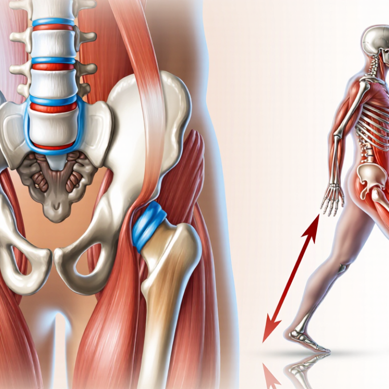
The hip is a ball-and-socket joint built for stability, movement, and weight transfer. The rounded femoral head fits inside the acetabulum, a cup-shaped part of the pelvis. Smooth cartilage covers both surfaces. It reduces friction as the hip bends, rotates, and bears body weight.

A deeper rim of fibrocartilage, called the labrum, helps seal the joint. The joint capsule and strong ligaments limit excessive motion without making the hip completely rigid. Around this structure, the gluteal muscles, hip flexors, adductors, and deep rotators create controlled movement. Their timing matters. A strong muscle may still produce inefficient motion when coordination is poor.

The femoral neck also affects leverage between the pelvis and thigh. Small differences in bone shape can change contact pressure and movement patterns. During walking, the hip transfers force from the ground through the leg into the pelvis. During a squat, the joint must manage compression while allowing forward flexion.

This model is useful, but incomplete. Real hips are not identical mechanical parts. Age, training history, previous injury, and daily habits can alter how forces move through the joint. In movement assessment, symptoms do not always reveal one clear structural cause. That uncertainty deserves attention, especially when a simple diagram seems too convincing.

What Is Hip Joint Biomechanics and Why Does It Matter?

Hip joint biomechanics describes how the femoral head, acetabulum, muscles, ligaments, and surrounding tissues work together to produce movement and transfer body loads. The hip is a ball-and-socket joint that supports activities such as walking, stair climbing, running, and rising from a chair.

The chart shows approximate normal hip range-of-motion values for healthy adults. Actual measurements vary with age, testing position, flexibility, and clinical technique. Adequate mobility and balanced muscle forces help maintain joint stability, efficient movement, and appropriate load distribution across the hip.

How Forces and Movements Act on the Hip

What Is Hip Joint Biomechanics and Why Does It Matter?

Hip biomechanics describes how forces and movements pass through the hip during daily activity. The joint must balance stability with mobility. Walking, climbing stairs, and standing on one leg create different mechanical demands.

During a step, the hip carries forces from the body above and the ground below. When the foot contacts the floor, the ground reaction force travels upward through the leg. Hip muscles then control the pelvis, preventing it from dropping toward the unsupported side. The gluteus medius is especially important here. Weakness or delayed muscle control may change walking patterns and increase stress around the joint.

Movement also depends on joint position. A deep squat increases hip flexion and may raise compressive forces, especially when the trunk leans forward. Turning the thigh inward or outward changes how the femoral head loads the socket.

Small changes can matter. Yet biomechanics is not a simple equation. Pain, strength, flexibility, previous injury, and movement habits interact differently in each person. A careful assessment should compare symptoms with observed motion, not rely on one posture or test. Even experienced clinicians can miss subtle coordination problems without repeated observation. That limitation deserves attention.

Biomechanics in Walking, Running, and Daily Activities

Hip joint biomechanics describes how the pelvis, thighbone, muscles, and connective tissues share movement and load. In walking, the hip manages repeated forces near 2.4 times body weight, according to established gait-analysis research. Each step also needs coordinated pelvic control. A small weakness in the side hip muscles can make the pelvis drop and increase stress around the knee or lower back.

Running raises the demand. Research commonly estimates hip joint forces near five times body weight during running, although speed, stride length, footwear, and surface change the result. The World Health Organization reported in 2022 that 31% of adults worldwide were insufficiently active. That matters because stronger movement habits can support capacity for stairs, lifting, and longer walks. Daily activities are not trivial. Standing from a chair requires hip extension, balance, and timing.

Form is not everything. Context matters more. A 2023 clinical review in the Journal of Orthopaedic & Sports Physical Therapy emphasized individualized load management for hip-related conditions. Pain does not always equal tissue damage, and painless movement is not automatically efficient. Clinicians should examine strength, range of motion, training volume, sleep, and recovery together. I find simple advice appealing, but “keep your knees aligned” can be too rigid. Bodies adapt differently. The better question is whether movement remains controlled, repeatable, and appropriate for the person’s current capacity.

Clinical Importance and Common Biomechanical Disorders

What Is Hip Joint Biomechanics and Why Does It Matter?

Hip biomechanics describes how the joint, muscles, bones, and pelvis share movement and load. Clinically, small changes can alter walking, stair climbing, and balance. Limited hip rotation may increase stress around the lower back or knee. Weak gluteal muscles can also reduce pelvic control during single-leg stance.

Common biomechanical disorders include hip osteoarthritis, femoroacetabular impingement, developmental dysplasia, labral injury, and gluteal tendinopathy. The World Health Organization reported that osteoarthritis affected 528 million people worldwide in 2019. About 60% were women. The Global Burden of Disease Study 2019 also identifies aging and obesity as major contributors to osteoarthritis disability. These figures matter in practice. Yet pain does not always equal structural damage. Our first explanation can be wrong. A careful assessment should combine symptoms, gait, strength, joint range, and imaging when appropriate.

Tips: Notice whether pain appears during sitting, pivoting, or walking uphill. Record the activity, duration, and pain level. Avoid forcing painful stretches. A physiotherapist or qualified clinician can assess movement patterns and determine whether weakness, stiffness, or joint structure is the main concern. Treatment should match the person, not only the scan. Even experienced clinicians must reconsider their assumptions when progress is slow.

What Is Hip Joint Biomechanics and Why Does It Matter? - Clinical Importance and Common Biomechanical Disorders

Biomechanical Dimension Typical Reference or Physiological Feature Clinical Importance Common Biomechanical Disorder or Alteration Potential Functional Effect Common Assessment Methods
Hip joint structure The femoral head fits into the acetabulum, forming a synovial ball-and-socket joint designed for mobility and load transfer. Joint geometry influences stability, contact pressure, range of motion, and the distribution of forces during standing and movement. Acetabular dysplasia, excessive acetabular coverage, femoroacetabular impingement, or structural deformity. Groin pain, restricted motion, labral stress, cartilage overload, or early degenerative change. Clinical examination, standing radiographs, and cross-sectional imaging when indicated.
Range of motion Hip motion includes flexion, extension, abduction, adduction, and internal and external rotation; normal values vary with age, sex, testing position, and measurement technique. Adequate mobility is required for walking, stair climbing, sitting, dressing, and squatting. Capsular stiffness, osteoarthritis, postoperative restriction, or impingement-related loss of flexion and internal rotation. Shortened stride, compensatory pelvic motion, difficulty putting on footwear, and painful gait. Goniometry, functional movement testing, and comparison with the opposite side.
Joint loading Hip joint reaction forces commonly exceed body weight during ordinary activities and rise further with running, stair climbing, or jumping. Load magnitude and direction affect cartilage health, bone adaptation, implant stresses, and symptom behavior. Excessive impact loading, altered force distribution, weak abductor control, or abnormal gait mechanics. Activity-related pain, fatigue, reduced walking tolerance, and increased tissue stress. Gait analysis, force-plate testing, instrumented activity assessment, and clinical load-response history.
Hip abductor function The gluteus medius and related muscles stabilize the pelvis during single-leg stance and control femoral motion in the frontal plane. Abductor strength and timing help maintain pelvic alignment and reduce excessive hip adduction. Abductor weakness, tendon pathology, pain inhibition, or superior gluteal nerve dysfunction. Contralateral pelvic drop, trunk lean, lateral hip pain, and reduced single-leg stability. Manual muscle testing, dynamometry, single-leg stance, step-down testing, and gait observation.
Femoral alignment Femoral version and neck-shaft geometry influence hip rotation, clearance, lever arms, and the direction of joint contact forces. Alignment affects movement efficiency and may modify the risk of impingement or instability. Femoral anteversion or retroversion, coxa vara, coxa valga, or rotational malalignment. In-toeing or out-toeing, limited rotation, altered gait, and uneven articular loading. Rotational profile examination, radiographs, and CT or MRI-based assessment when clinically justified.
Pelvic control The pelvis coordinates trunk and lower-limb movement and transfers forces between the spine and hips. Pelvic position changes the functional orientation of the acetabulum and influences hip stability and range of motion. Excessive anterior or posterior pelvic tilt, pelvic obliquity, or poor lumbopelvic coordination. Increased hip flexor or extensor demand, lumbar compensation, and altered gait mechanics. Postural assessment, lumbopelvic movement testing, gait observation, and functional tasks.
Gait mechanics Normal walking requires coordinated hip extension, controlled flexion, pelvic rotation, and stable single-limb support. Gait analysis identifies compensations that may not be obvious during non-weight-bearing examination. Antalgic gait, Trendelenburg gait, reduced hip extension, or excessive hip adduction. Lower walking speed, asymmetrical step length, increased energy expenditure, and secondary knee or back symptoms. Observational gait assessment, video analysis, wearable sensors, or three-dimensional motion analysis.
Femoroacetabular clearance The femoral head and neck should move through functional ranges without damaging contact between the femur and acetabular rim. Clearance is important for identifying motion-related pain and distinguishing impingement patterns from other causes of hip symptoms. Cam morphology, pincer morphology, or combined femoroacetabular impingement. Pain with flexion and rotation, reduced internal rotation, labral injury, and cartilage damage. Provocative clinical tests, radiographs, and MRI or MR arthrography when appropriate.
Load distribution and cartilage health Articular cartilage distributes compressive forces and enables low-friction movement across the hip joint. Balanced loading helps preserve joint surfaces and supports efficient movement. Osteoarthritis, cartilage loss, labral pathology, or chronic focal overload. Stiffness, crepitus, reduced activity, pain during weight-bearing, and progressive functional limitation. History and physical examination, radiographs for structural change, and MRI for soft-tissue or cartilage assessment.
Clinical rehabilitation target Effective treatment aims to improve capacity, movement control, strength, mobility, and tolerance to progressively increased loads. Biomechanical analysis supports individualized exercise selection and helps monitor functional progress. Persistent movement compensation, deconditioning, fear of movement, or inadequate load progression. Delayed recovery, recurrent symptoms, reduced participation, and dependence on compensatory strategies. Patient-reported outcomes, strength testing, range-of-motion reassessment, functional tests, and activity monitoring.

Note: Reference findings vary according to age, body structure, activity level, symptoms, testing position, and measurement method. Biomechanical findings should be interpreted together with the clinical history and physical examination.